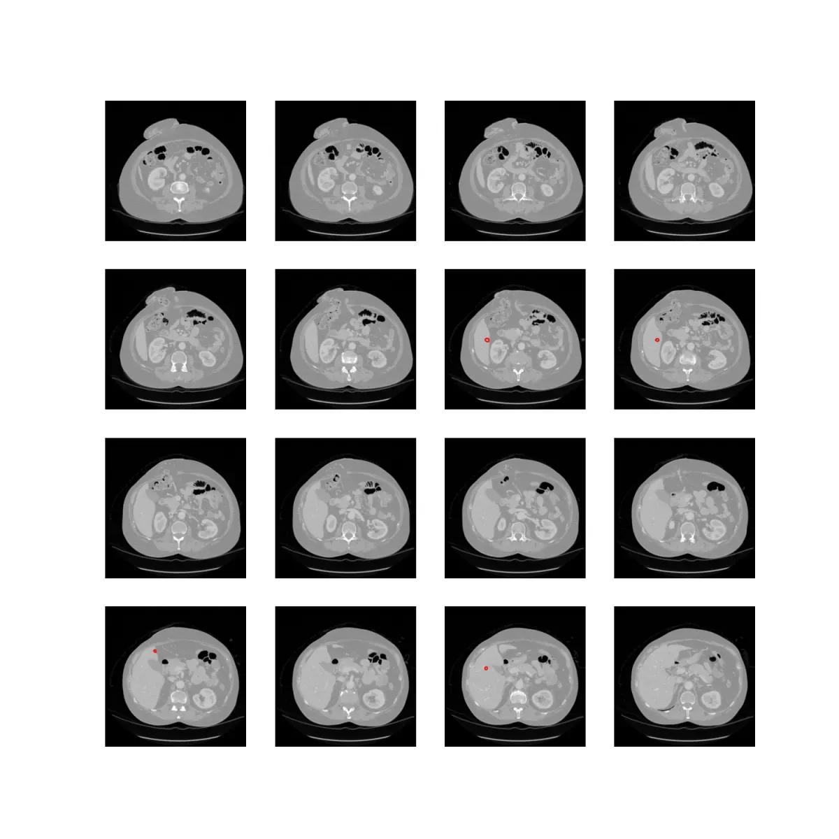
CT · PET/CT · MRI
LymphNode AI
Detect, segment, and measure lymph nodes across the body — for staging and follow-up.
510(k) pathway in progress
OncoAI Suite · AI-Native Oncology Imaging Suite
Six AI engines under a single viewer: BrainTumor in clinical use today, LymphNode on the 510(k) pathway, LungNodule in clinical research use, and kidney / liver / whole-body PET/CT in development. CT, MRI, and PET/CT — same UI, same pipeline.
The suite
CT · PET/CT · MRI
Detect, segment, and measure lymph nodes across the body — for staging and follow-up.
510(k) pathway in progress
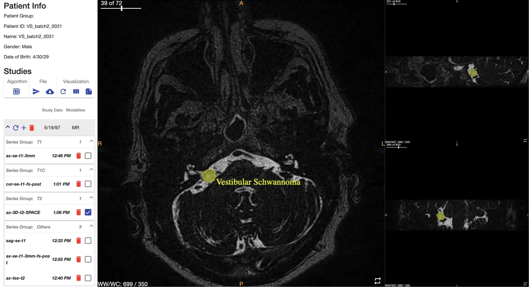
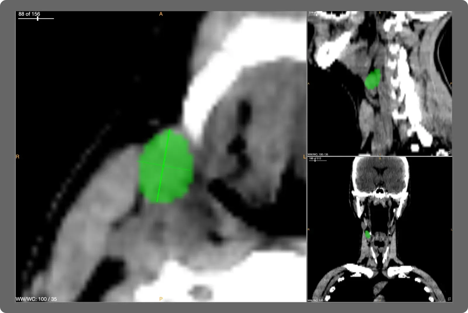
Multi-parametric MRI
Glioma, brain metastasis, and vestibular schwannoma — segmented with volumetric tracking across visits.
Validated · in clinical use
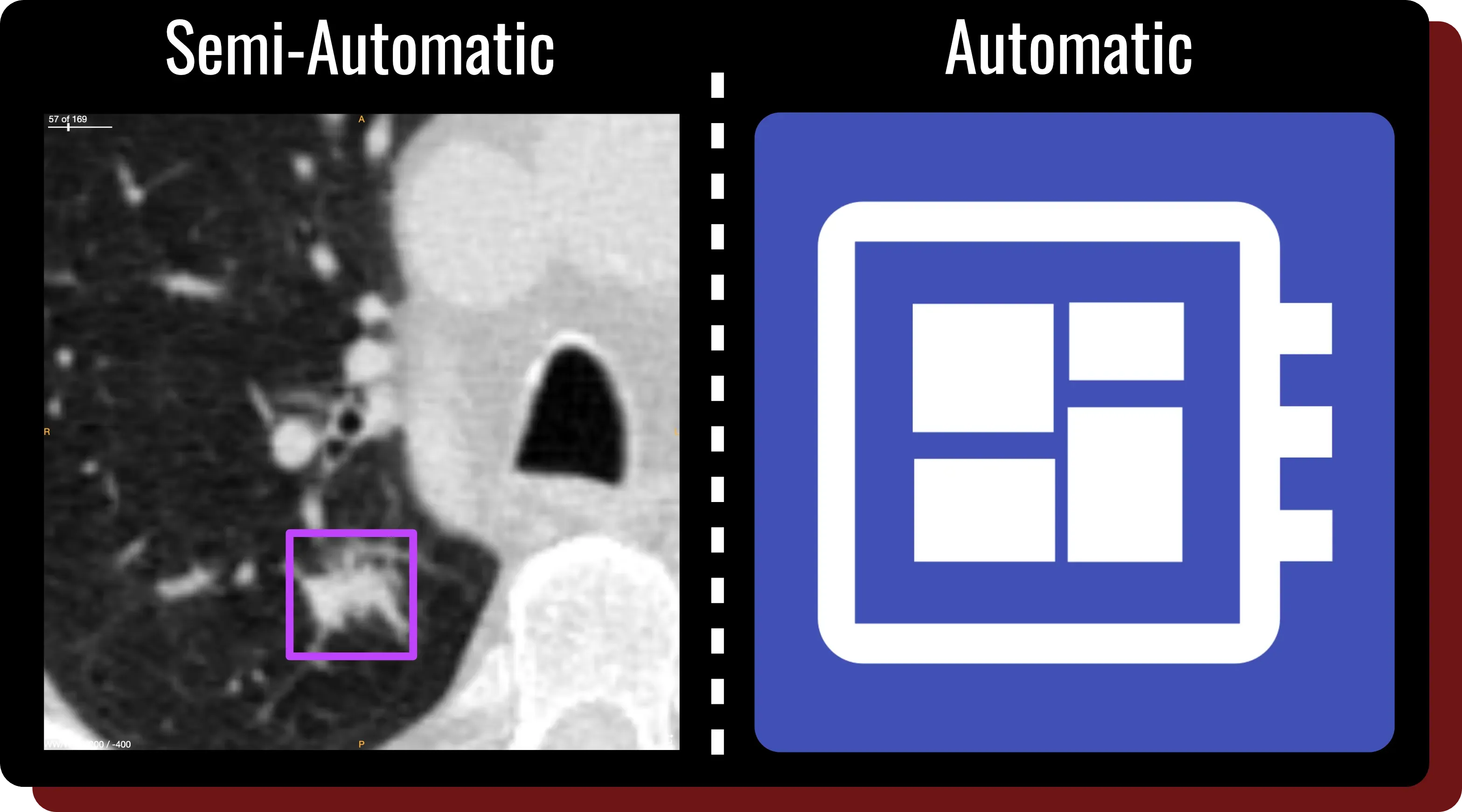
Chest CT
Detection, segmentation, and Lung-RADS scoring on chest CT — semi-automatic or fully automatic.
In clinical research use
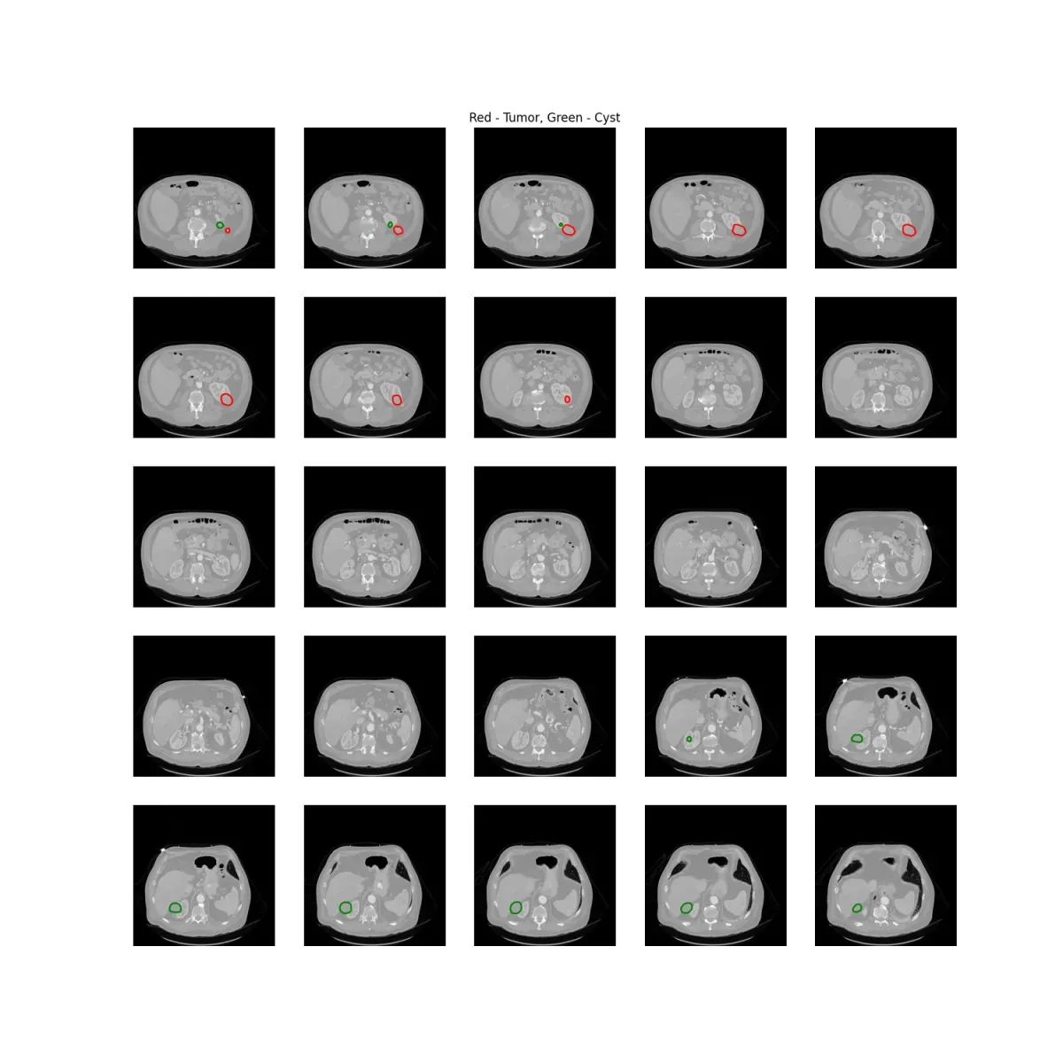
CT (contrast)
Automatic segmentation of renal tumors and cysts — for staging, surveillance, and surgical planning.
In development
CT (contrast)
Automatic segmentation of liver tumors from contrast-enhanced CT for response assessment.
In development
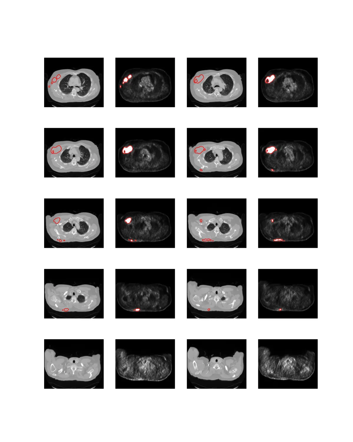
PET/CT
Whole-body lesion segmentation on PET/CT — coregistered against CT anatomy.
In development
How it works
Cancer targets are small, low-contrast, and unpredictably located. A single segmentation model misses them or floods the report with false positives. OncoAI separates the problem into three networks — each tuned to a different objective — and the LymphNode model is the most-validated example.
01
RetinaUNet3D scans patch-by-patch to surface every candidate. Recall is prioritized so nothing is missed downstream.
02
EfficientNetV2S with 3-panel augmentation rules out false positives without sacrificing the sensitivity from stage 1.
03
UNet3D produces masks for each confirmed finding. Long/short-axis and volume measurements come directly from the masks.
666
Scans · LymphNode validation
625 CT + 41 MRI — head & neck, thorax, abdomen, pelvis. Train 444 / val 222.
80%
True-positive rate (distance-hit)
79.93% sensitivity at 1.27 false positives per true positive on the 222-scan validation set.
0.69
Dice (node-level segmentation)
0.6922 ± 0.2283 mean ± SD on 212 scans with ground-truth masks.
Publications
On-premise web service · DICOM C-FIND / push / upload · NIfTI / NRRD · DICOM Seg / CSV / report out · Batch CLI for cohort runs.